1 minute read
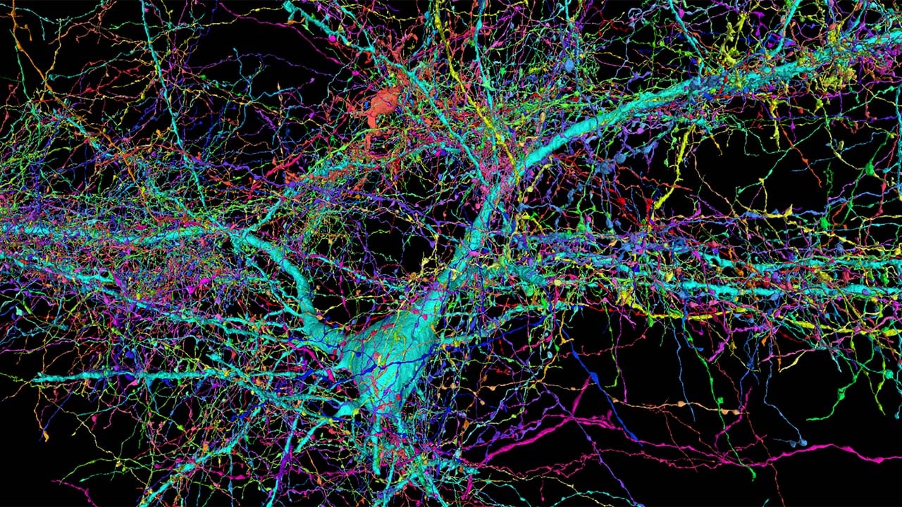
Harvard researchers, along with Google Research, have produced incredible new 'synaptic-level' images of a fragment of the human brain, showing previously unseen levels of detail.
The images were produced from a tiny fragment of human brain tissue; one cubic millimetre to be precise. That might seem minuscule, but even that tiny fragment contains 57,000 cells, 230 millimetres of blood vessels, and 150 million synapses. The new research has produced the largest ever dataset of neural connections to date.
The project, which was headed up by Jeff Lichtman, the Jeremy R. Knowles Professor of Molecular and Cellular Biology, took a decade to complete, and aimed to combine Lichtman’s electron microscopy imaging with Google's AI algorithms to create a colour coded 3D map of a mammal's brain, right down to a cellular level.
The fragment, which was taken from a 45 year old woman undergoing a procedure to treat epilepsy, was sliced into 5000 slices, each 34 nanometers thick, and then imaged using electron microscopy. The resulting data from this latest research produced an astounding 1,400 terabytes of data.

To put that into further context, in order to map an entire human brain would require over a billion terabytes (a zettabyte). The project's ultimate goal is to produce a full map of the brain of a mouse, which while much smaller than a human, would still require 1000 times the data of the single fragment map.
The new dataset contains details that have never been seen before, including a rare but powerful set of axons connected by up to 50 synapses. The researchers also discovered oddities, such as axons that formed extensive whorls. Although it's not clear if these were as a result of the patient's epilepsy.
The full dataset is publicly available, and you can see some examples here, although load times can be extensive. Google collaborator, Viren Jain, said "“Given the enormous investment put into this project, it was important to present the results in a way that anybody else can now go and benefit from them,”
Research like this will be vital in the future for understanding different brain conditions and diseases, but it is clear that there is a mountain to climb before even a small mammal's brain can be fully mapped.
Tags: Science

RedShark 2020 @ All rights reserved.

Comments